CURRENT
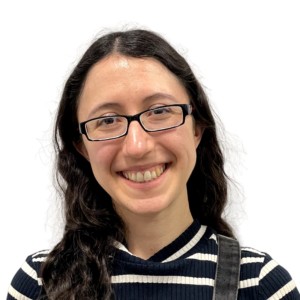
Eva’s thoughts on the program…
During my PhD, I used optical microscopy to probe different topics in cell biology ranging from early human placental development to CAR T-cell signaling. Through this work, I not only discovered my own passion for microscopy, but also how much I enjoyed explaining microscopy fundamentals to my colleagues in a way that made them excited about it too! Since starting as an Advanced Microscopy Fellow at HMS in 2023, I have had the opportunity to broaden my foundational understanding of microscopy, as well as my teaching and core management skills, under the mentorship of the absolutely brilliant CITE team — and I have loved every second of it! The fellowship program is the perfect opportunity to explore what it is like to work at a core facility and actively contribute educational resources to the scientific community. I have thoroughly enjoyed delving deeper into microscope maintenance essentials and strengthening my microscope troubleshooting skills. However, I have particularly enjoyed helping the team empower the myriad of HMS researchers successfully implement microscopy in their science through training sessions and workshop programming. Working with Jennifer, Anna, Talley, and Federico is so fun not only because they love all things microscopy, but also because they deeply care about sharing useful resources and tools for the broader community. I am honored to have the opportunity to contribute to their existing programming and new exciting projects on the horizon (like Microtutor!), and hope to explore more ways to help empower researchers using microscopy through my independent fellow projects.
This program is truly unique and special – I highly recommend it to anyone interested in pursuing a career in microscopy +/- core management!
ALUMNI
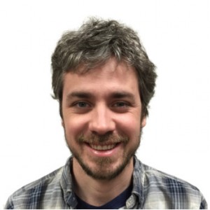
Talley’s thoughts on his experience with the fellowship program:
During my postdoc, I attended Jennifer’s Quantitative Imaging course at Cold Spring Harbor. There I discovered my passion for all things microscopy, and applied for the Advanced Microscopy Fellowship program at HMS the following year. During the fellowship, I was able to immerse myself in microscopy: forming a solid theoretical foundation in quantitative imaging, as well as a breadth of practical techniques including live-cell imaging, FRET, FRAP, confocal, TIRF, and super-resolution microscopy. In addition, I learned a lot about the day-to-day operations and considerations when running a very heavily used imaging core facility. I was particularly involved in optimizing acquisition and reconstruction protocols for structured illumination microscopy. Additionally, I developed multiple lectures for on-campus workshops and our course at Cold Spring Harbor.
Starting in 2015, I accepted a position as a Research Associate in the Cell Biology Department at HMS, where I manage a small core specializing in advanced optical imaging techniques including 3D-Structured Illumination super-resolution microscopy, as well as two light sheet microscopes: a diSPIM, and a Lattice light sheet microscope. Jennifer’s post-doctoral fellowship was an incredible experience that provided me with more resources than I could have hoped for to feed my desire to learn more about microscopy and core facility management.
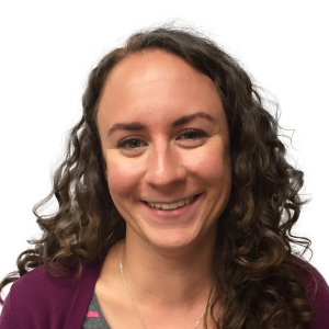
Anna’s thoughts on her experience with the fellowship program:
The Microfellows program gave me the opportunity to learn about all aspects of core facility life, from user training and consultation, to microscope maintenance, to grant writing and management. On the technical side, I’ve learned the theory and practice of a wide variety of advanced microscopy techniques, including FRAP, ratiometric imaging, single-molecule imaging, SIM, and STORM. I’ve tested, reinforced, and deepened my understanding by training users in each of these techniques.
I love teaching, so I spent a lot of my time as a fellow working on our microscopy workshop program at HMS. I lectured in our workshops, created new lectures and lab exercises, and coordinated workshop logistics. When I want to learn more about a topic, I develop a lecture about it; there’s always an enthusiastic and appreciative audience for microscopy lectures at HMS. I’m also very much involved in our annual Cold Spring Harbor course, and in the months leading up to the course, I help to write lab exercises, prepare samples, and coordinate equipment.
In addition to teaching, I worked on several other projects as a fellow, including automation of quarterly microscope inspections, methods to measure phototoxicity, and a review on validation and bias in microscopy experiments.
In 2019, I accepted a position as Associate Director of Imaging Education in the NIC@HMS. In my new role, I’ll build on my work as a fellow by continuing to expand our workshop program and develop new microscopy education offerings for HMS and beyond. My time as a fellow was incredibly rewarding and productive, and I’m looking forward to sharing that experience with the next generation of Microfellows.
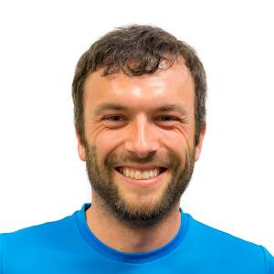
Michael’s thoughts on his experience with the fellowship program:
In March 2016, I started working as an Advanced Microscopy Fellow in Jennifer’s core facility, which is located on a historic, yet international and diverse campus that hosts numerous renowned researchers. From the outset, I was profiting from the team’s extensive expertise in quantitative light microscopy and teaching – and their willingness to share that knowledge. Thanks to Jennifer’s unique fellowship program, I significantly expanded my expertise in these areas and learned advanced techniques such as spinning disc confocal and total internal reflection microscopy to great detail. Teaching was a heavy focus of my work. Not only did I train and advise researchers in the use of advanced microscopy techniques, I also gave lectures, taught workshops and contributed to our famous yearly “Quantitative Imaging” course in Cold Spring Harbor. Furthermore, I truly appreciated the freedom of pursuing my own projects. This included designing and building a compact light sheet microscope for research and education, designing and integrating four new Nikon widefield microscopes for various applications, as well as writing scientific reviews and book chapters.
In December 2018, I finished my postdoc position in Jennifer’s facility. In January 2019, I took on a new role as Field Application Specialist for the invol3D Flamingo project (https://involv3d.org/flamingo). There, I’ll collaborate with researchers using traveling, powerful light sheet microscopes. Throughout the process of transitioning into my new job, Jennifer was – and continues to be – very supportive. The hard and soft skills I acquired during my time in her facility, as well as the academia and industry connections I built there, will be of tremendous help in my new role.
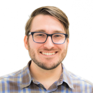
George’s thoughts on his experience with the fellowship program:
During my Ph.D. research, I used light microscopy and image analysis to answer questions about the cell biology of neurons. Learning about microscopy and helping other researchers with that knowledge were my favorite aspects of the Ph.D. experience.
The Advanced Microscopy Fellowship program allowed me to use my research experience to advise HMS researchers in their diverse light microscopy experiments. By assisting researchers with a variety of experimental setups, the breadth and depth of my understanding expanded beyond what was required of my Ph.D. research. Using theoretical microscopy knowledge to better train, advise, and teach researchers was a wonderful challenge.
In December 2019, I finished the Advanced Microscopy Fellowship program. In January 2020, I took on a new role as a Senior Research Technologist in the Cell & Tissue Imaging Center at St. Jude Children’s Research Hospital in Memphis, TN. My experience as a Microfellow with Jennifer has helped me to transition from being an experimental biologist who fancies microscopes to becoming an experimental microscopist who fancies biological experiments.
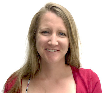
Rylie’s thoughts on the program:
I started my fellowship in the NIC in summer 2019, after finishing my PhD. Microscopy has always been my favorite part of being a scientist, and my time as a fellow vastly deepened my theoretical and practical understanding of quantitative microscopy, from the basics to advanced techniques. I am amazed by how much I learned from Jennifer and the rest of Team NIC throughout my time there. I also benefited from the NIC’s large and diverse user base, which gave me experience advising and training scientists in a broad range of modalities and applications. During my fellowship my primary interests were structured illumination microscopy (SIM) and microscopy education. Working with Anna on the NIC workshop program, I discovered a passion for teaching microscopy, and was able to gain experience developing lectures, design workshops & tutorials, lead discussion groups and help plan and execute an online lecture series. Serving as a TA in Jennifer’s Quantitative Imaging course at Cold Spring Harbor Laboratory was the most rewarding (and the most fun) experience of my career so far.
My fellowship taught me that I want microscopy education to be central to my future career. After leaving the NIC, I accepted a new role as a Staff Scientist at the Marine Biological Laboratory, where I will split my time between assisting and training scientists in the use of advanced home-built instruments, and expanding the MBL’s year-round microscopy education program. I am sure that the skills that I learned from Team NIC in core management, user training and teaching microscopy will be of great use to me in this position. Overall, this fellowship was an incredible experience that gave me resources and opportunities that I strongly believe I would not have been able to obtain otherwise.
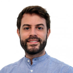
Federico’s thoughts on the program:
I started my fellowship in the NIC in summer 2019, after finishing my PhD. Microscopy has always been my favorite part of being a scientist, and my time as a fellow vastly deepened my theoretical and practical understanding of quantitative microscopy, from the basics to advanced techniques. I am amazed by how much I learned from Jennifer and the rest of Team NIC throughout my time there. I also benefited from the NIC’s large and diverse user base, which gave me experience advising and training scientists in a broad range of modalities and applications. During my fellowship my primary interests were structured illumination microscopy (SIM) and microscopy education. Working with Anna on the NIC workshop program, I discovered a passion for teaching microscopy, and was able to gain experience developing lectures, design workshops & tutorials, lead discussion groups and help plan and execute an online lecture series. Serving as a TA in Jennifer’s Quantitative Imaging course at Cold Spring Harbor Laboratory was the most rewarding (and the most fun) experience of my career so far.
My fellowship taught me that I want microscopy education to be central to my future career. After leaving the NIC, I accepted a new role as a Staff Scientist at the Marine Biological Laboratory, where I will split my time between assisting and training scientists in the use of advanced home-built instruments, and expanding the MBL’s year-round microscopy education program. I am sure that the skills that I learned from Team NIC in core management, user training and teaching microscopy will be of great use to me in this position. Overall, this fellowship was an incredible experience that gave me resources and opportunities that I strongly believe I would not have been able to obtain otherwise.

